From phenotype to genotype
Publié le 06 oct 2023Lecture 3 min
Prenatal Diagnosis of Left Atrial Isomerism
ABSTRACT
Situs solitus is defined as normal development and positioning of the thoracic and abdominal organs.
Heterotaxy syndromes describe a spectrum of abnormal organ location including conditions such as asplenia and polysplenia and are best classified by the morphology of the cardiac atria as right and left isomerism. In addition, heterotaxy syndromes are present in about 2-24% of children with congenital heart defects (CHD). A 31-year old G2P1 caucasian woman presented at 13 weeks of gestation for routine first trimester assessment.
The stomach was present but located centrally rather than on the left side of the abdominal cavity and the cardiac axis deviated to the left. At 17 weeks, the left superior vena cava and the aorta had obviously smaller diameter than the pulmonary artery. Fetal karyotype was normal (46XY), but Whole Exome Sequencing detected two missense variants of uncertain significance in the gene PKD1L1.
The infant was born by cesarean section at 38 weeks and the diagnosis was confirmed after birth. Left isomerism is characterized by “double” left structures and underdevelopment or absence of right structures. The prognosis depends mainly on the severity of the heart defects and the development of hydrops.
INTRODUCTION
Situs solitus is defined as normal development and positioning of the thoracic and abdominal organs. Heterotaxy syndromes describe a spectrum of abnormal organ location including conditions such as asplenia and polysplenia and are best classified by the morphology of the cardiac atria as right and left isomerism(1).
CASE REPORT
A 31-year old G2P1 caucasian woman presented at 13 weeks of gestation for routine first trimester assessment. Her family and medical history were unremarkable and there was no consanguinity.
Her first pregnancy resulted in a healthy term infant. The fetus was normally grown (CRL=68.5 mm) and nuchal translucency was 2.4 mm.
The stomach was present but located centrally rather than on the left side of the abdominal cavity (Figure 1). In addition the cardiac axis deviated to the left. Maternal biochemistry was normal (βhCG: 0.308 MoM, PAPPA: 0.518 MoM) and the risk for the common trisomies was low (1 in 11,941). Repeat ultrasound examination at 17 weeks showed that the stomach remained in central position, there was persistent left superior vena cava and the aorta had obviously smaller diameter than the pulmonary artery (Figures 2, 3). The results were confirmed by fetal echocardiography.Amniocentesis was performed at the request of the parents; the fetal karyotype was normal (46XY) and there were no additional findings from CGH arrays analysis. Fetal MRI at 24 weeks confirmed the central position of the stomach whereas no structural anomaly was detected. Repeat fetal echocardiography at 29 weeks showed normal growth of the aorta and the aortic arch, two morphologically left atria and interrupted inferior vena cava thus establishing the diagnosis of left atrial isomerism. Further investigations by Whole Exome Sequencing detected two missense variants of uncertain significance in the gene PKD1L1. Pathological mutations with heterozygosity in this gene have been associated with autosomal visceral heterotaxy. The parents were counseled by a clinical geneticist on the favorable prognosis in the absence of a heart defect and decided to continue the pregnancy. Repeat MRI scan at 35 weeks identified multiple small spleens and malrotation of the bowel.The infant was born by cesarean section at 38 weeks (birth weight 3,300 gr). The diagnosis was confirmed after birth. He is currently discharged at home and is scheduled for bowel surgery.
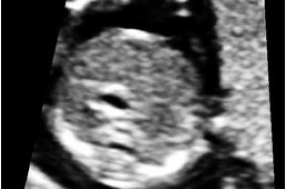
Fig. 1: Cross-sectional view of the fetal abdomen at
13 gestational weeks with central positioning of the stomach
Fig. 2: Cross-sectional view of the fetal abdomen at
17 gestational weeks with the stomach positioned centrally.
Fig. 3: Cross-sectional view of the thorax depicting the three vessel view.
Note the smaller diameter of the aorta and the persistent left superior vena carva (white arrow).
DISCUSSION
Heterotaxy syndromes are present in about 2-24% of children with congenital heart defects (CHD). Left isomerism is more common. The condition is genetically diverse including autosomal dominant, recessive and X-linked inheritance(2). The recurrence risk in subsequent pregnancies is estimated to be about 10%.Left isomerism is characterized by “double” left structures and underdevelopment or absence of right structures. Usually there is absence of a morphologically right atrium and the sinus node, leading to bradyarrhythmia of complete heart block (up to 70% of the cases). The cardiac axis is often deviated centrally or to the left, the left superior vena cava may be present and the pulmonary veins may be abnormally connected(3). Hydrops may develop in about one third of cases. Other anomalies include polysplenia, right-sided stomach, anomalies of the upper gastro-intestinal tract, left-sided or medial liver and absent gallbladder(4). The prognosis depends mainly on the severity of the heart defects and the development of hydrops.
CONCLUSION
Detailed examination of the fetal anatomy at the 11-13 weeks scan can raise suspicion for the presence of rare genetic conditions and guide timely and appropriate investigations(5).
Attention, pour des raisons réglementaires ce site est réservé aux professionnels de santé.
pour voir la suite, inscrivez-vous gratuitement.
Si vous êtes déjà inscrit,
connectez vous :
Si vous n'êtes pas encore inscrit au site,
inscrivez-vous gratuitement :
