Fetal imaging
Research
Publié le 10 Mar 2019Lecture 8 min
New 3D embryo-foetal imaging using immunostaining
Presented by Gérard COULY, Work from the Institut de la Vision [Vision Institute], Inserm UMRS 968, Paris
Clarifying human development using 3D imaging of transparent whole embryos and organs
Generating a precise cellular and molecular cartography of the human embryo is essential to understand the mechanisms of organogenesis in normal and pathological conditions. Here, we combined whole-mount immunostaining, 3DISCO clearing and light-sheet imaging to start building a three-dimensional cellular map of human development during the first trimester of gestation. We provide 3D images at an unprecedented resolution of the developing peripheral nervous system, vascular system, cardiopulmonary system, urogenital system and muscular system. We show that the adult-like pattern of skin innervation is established before the end of the first trimester with important intra- and inter-individual variations in nerve branches. We also present evidence for a differential vascularization of male and female genital tracts concomitant to sex determination. This work paves the way for a cellular and molecular reference atlas of human cells, which is critical to understanding human developmental disorders.
Introduction
The Institut de la Vision work group, led by Alain Chedotal (UCPM, Inserm UMRS 968, Paris 6 Sorbonne), published the imaging of numerous developing organ structures from embryos and foetuses, shown using immunostaining, from the end of the first trimester to the beginning of the second, between the 6th and 12th week after conception or the 8th to 14th week of amenorrhoea in the review Cell in 2017(1).
The images, obtained in high definition, then reconstructed in 3D, enabled the creation of cellular and molecular mapping and development of the human embryo and becoming an early foetus to be documented.
All imaging from the article was supplemented by the freely accessible website containing 80 videos: http://www.transparent-human-embryo.com/.
The new morphological analysis using immunostaining sheds light on developmental anatomy over a period that, until now, has been poorly documented, i.e. at the end of organogenesis, and constitutes a preliminary and essential step towards the normal and pathological organ recognition looked for during antenatal ultrasound.
Material and methods(1)
The thirty six embryos (n = 7) and foetuses (n = 29) studied and analysed were collected by institutions that ensured statutory obligations such as consent, ethics, samples, embryonic and foetal fixations, and the collection of essential genomic and phenotypic data.
The ages and sexes of the specimens were determined before fixation and the specimens were treated in immunohistochemistry with primary and secondary antibodies, totalling 19, targeted at specific epitopes of cells with phenotypes that were already expressed. Amongst the antibodies used for immunostaining were:
– peripherin, targeted at the protein in the filaments from neurones in the sensitive sensory peripheral nervous system ;
– Tag1, targeted at the sensitive sensory nerve fibres ;
– anti-myosins of striated muscle and anti-myosins of smooth muscle, targeted at the smooth musculature of the vascular walls ;
– choline acetyltransferase, recognising the motor nerves ;
– antibodies against the SOX10 and SOX2 proteins, for recognition of Schwann cells in the sensory nerves ;
– antibodies targeted at the PLVAP protein, marking the vascular endothelia.
The immunostained developing essential organ cells were identified in this way in relation to their phenotypes.
The images were acquired using light sheet fluorescence ultramicroscopy, working stratigraphically in virtual cross sections of 3 microns, and reconstructed in 3D, thus enabling permanent preservation of the sample.
This procedure allowed the organ volumes, but also the nerve, muscle and internal vessel structures of the organs, to be visualised at an early stage, together with cellular organisation at high resolution where possible.
The following developing embryo and foetal areas were explored and visualised in 3D :
– the cardiopulmonary system ;
– the peripheral nervous system ;
– the central nervous system ;
– the somatic muscles ;
– the urogenital tract ;
– the vascular system ;
– the digestive tract.
Results
The imaging obtained in high resolution relates to the development of numerous organs and organ systems.
The earliness of phenotypic and anatomical determination of the organs
This imaging revealed the early nature of establishing cellular phenotypes and organ constructions that took place from the end of the first gestational trimester. These appeared to us as miniatures or reduced models of future organs that we will see in adults. We do not currently know the molecular factors that control embryo-foetal organ growth from the end of organogenesis. However, this early organogenetic nature could have as application and justify the possibility of early diagnosis of numerous dysmorphologies, from the end of the first trimester to the 12th week after amenorrhoea. This is particularly demonstrative for the embryo's heart and body and its extremities (figures 1, 2, 3, 4 and 5).
Figure 1 : Embryo-foetal heart at 9 weeks after conception, 11th week of amenorrhoea. Its anatomy, both external as well as internal, vascular and nerve, is fully established. Note the scale. At this stage, the heart measures 3,000 microns x 3,000 microns x 4,000 microns.
Figure 2 : Human embryo body at 8 weeks after conception, 10th week of amenorrhoea. Left preparation, posterior view ¾ left, view of the posterior brain (brainstem and 4th ventricle), spinal cord and nerve emergences, destined for the head, chest and arms. The right preparation shows the upper half of the body (skeleton and muscles).
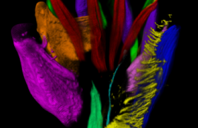
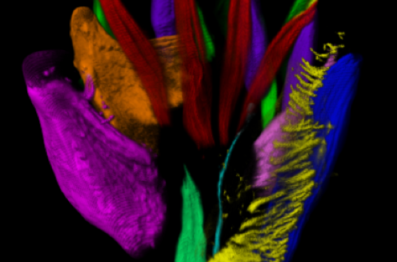
Figure 3 : Left hand at 9.5 weeks after conception. All the thenar and hypothenar musculature, as well as the interosseous and lumbrical muscles in dorsal and ventral views, were visualised and identified according to the different colour (immunostaining with anti-myosin).
Figure 4 : Left foot at 9.5 weeks after conception and its nerves, in immunostaining with choline acetyltransferase and Tag1, revealing the motor and sensory nerves in 3D.
Figure 5 : The brainstem and its nerves at 7.5 weeks after conception. Head and neck profile of the human embryo, showing cranial nerves III to XII from the brainstem, and the cervical roots destined for the face and neck with immunostaining with Tag1 and peripherin in 3D. Note the early nature and nerve density in the orofacial and cervical regions of the embryonic outline at this stage. The embryo becomes a foetus at 10 weeks after conception (12th week of amenorrhoea) and begins to suck and swallow thanks to the neurological organisation of the brainstem and its nerves. In box: the embryo face on at the same age.
Organogenetic earliness and functional embryo-foetal activity
The early nature of completed organogenesis and the density of the peripheral nervous system, particularly regarding the brainstem nerves (III > XII) and their oral, lingual and pharyngo-laryngeal sensory ganglia (figure 5) around 10 weeks after conception (12th week of amenorrhoea) correspond with the initial manifestations of the embryo-foetal oral development of sucking and swallowing shown in the ultrasound. In this way, these oral behavioural sequences, if fundamental for the future of the foetus and neonate, do not echo the joint and chronological development of the region of the central nervous system, which is in charge of neurological control (figure 4). Likewise, in this oral function context, the sensitive sensory and motor innervation of the tongue and the face has the same neurological maturity (figures 5, 6, 7 and 8).
Figure 6 : Brain and brachial-thoracic innervation at 8 weeks after conception, face on, using immunostaining with anti-myosin and Tag1 (I: olfactory nerve; II: optic nerve).
Figure 7 : Embryo tongue at 9.5 weeks after conception (views from behind, face on and in profile). Immunostaining using choline acetyltransferase and Tag1 shows the density of the motor nerves (XII), sensory nerves for general sensation (V, IX) and sensory gustatory nerves (VII bis, and IX) in 3D.
Figure 8 : Trigeminal innervation of the foetal face at 8 weeks after conception. Human embryo face at 8 weeks after conception. Immunostaining using Tag1 and anti-myosin reveals the sensory neurological density of the right and left trigeminal nerves, as well as the right and left oculomotor muscles in 3D (in red).
Asymmetries and variations
Amongst the data obtained, those collected before the end of the first gestational trimester, such as early embryo-foetal peripheral innervation of the limbs and their extremities, appeared to us to be similar to those of an adult; however, they already have significant asymmetries and intra- and interindividual variations(1).
The olfactory and optic nerves
The olfactory (I) and optic nerves, together with the chiasm (II), were well visualised at 8 weeks after conception, i.e. 10th week of amenorrhoea, as well as the oculomotor muscles (figures 8, 9 and 10).
Figure 9 : Presentation of the content of the eye socket at 8 weeks after conception, with the oculomotor muscles (in red) and optic nerve II (arrow, in green). I: olfactory nerve across the lamina cribrosa and in the embryonic nasal cavity.
Figure 10 : The optic chiasm at 8 weeks after conception with immunostaining in high definition, using Tag1, in 3D.
Sexual differentiation
Genital male or female differentiation and determination of sex appeared to have vascular origin.
Discussion
Cell differentiation of genetic origin during development is the result of the actuation of a cascade of transcription factors. It determines varied cell phenotypes, necessary for the development of organs with specific functions and therefore organisms. These identified transcription factors are the new analysis and comprehension tools for the study of morphogenesis of the human embryo.
Nineteen antibodies were used, targeted at these transcription factors, to monitor the developments of various organ systems visualised and studied using immunostaining.
This new imagery seemed to us to be an undeniable advancement in the analysis of organogenesis and the dynamics of embryo-foetal anatomy between the 6th and 12th week after conception, i.e. between the 8th and 14th week of amenorrhoea, a period of development that remains inadequately documented, particularly as this period relates to the end of organogenesis and the boundary between the embryonic and foetal stages.
The samples studied were immortalised using this analysis method. This method once again responds to the adage by Prof. André Delmas that "Death is useful to life".
The foetus sonographers can consult the website freely to make use of the organogenetic atlas covering the end of the embryonic period to the start of the foetal period, a period when certain functions, such as oral development, sucking and swallowing begin.
Visualisation of the nerve, vessel, muscle and intimate organ architectures (such as the lungs, for example) will provide essential completion for sonographic imagery, enabling earlier and earlier organ investigations.
Unfortunately, this high-definition progressive imagery, useful for visualising early embryo-foetal development, cannot yet be carried out in vivo using immunostaining. We believe that it will be in the future, when foetal-maternal chemistry and pharmacology have resolved the toxicity of antibodies and their harmful effects, allowing them to be used with no risk to the mother or foetus.
Numerous embryo-foetal structures are therefore constructed in the early 12 weeks of amenorrhoea stage, in a complete and final manner, but on a very reduced scale (the organs are several millimetres). This would allow early dysmorphological investigations to be carried out around the 12th week of amenorrhoea in the future, once the stated potential toxicity of the antibodies used is resolved.
This option of early and precise embryo-foetal dysmorphological investigation relating to anatomical structures such as nerves (optic chiasm, vestibulocochlear structures, etc.) or all other structures from warning signs onwards, which would need to be defined, could trigger the MTP decision at the age and stage when terminations are carried out. This would be congruent in terms of time with terminations and MTP.
Conclusion
We present the original imagery obtained from cellular and molecular mapping of the human embryo and foetus from the first trimester of pregnancy. It is essential to understand the normal and pathological mechanisms of organogenesis. The method consists of identifying the organs and their constituent tissues thanks to their transcription factors, which guaranteed cell differentiation, using specific molecular immunohistochemistry, that is revealed by 3D imaging. The progressive cellular and organ mapping obtained relates to the following development :
– peripheral and central nerve ;
– cardio-pulmonary and vascular ;
– urogenital ;
– muscular and limbs.
Each of the constituent tissues is located using immunostaining, thanks to the antibodies produced against the specific molecular cell targets.
New morphological information has been collected, such as the significantly early nature of the development of organs, the scale of which is considerably reduced, the establishment of nerve metamerics and cutaneous territories, or the vascular origin of sexual differentiation.
This work is a precursor to the early embryo-foetal imaging of the future. But for now, it remains much further upstream of foetal ultrasound, awaiting foetal-maternal pharmacological progress enabling the use of these antibodies in vivo without any risk.
Attention, pour des raisons réglementaires ce site est réservé aux professionnels de santé.
pour voir la suite, inscrivez-vous gratuitement.
Si vous êtes déjà inscrit,
connectez vous :
Si vous n'êtes pas encore inscrit au site,
inscrivez-vous gratuitement :